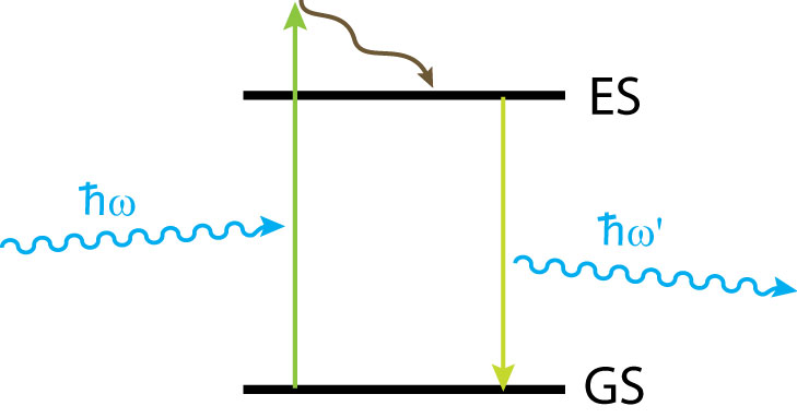
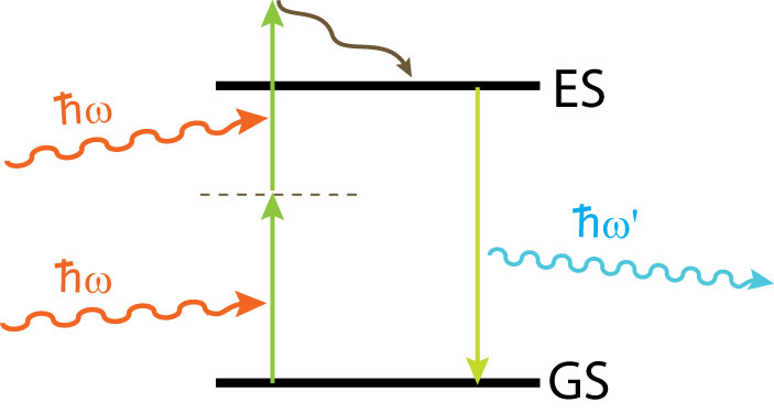
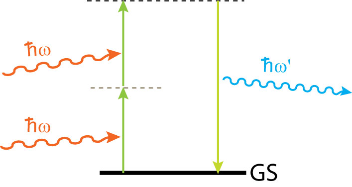
Members: Chih-Yu Jao In standard fluorescence microscopy, a sample is stained with a dye and then imaged by scanning a focused laser beam across the sample. The laser wavelength is chosen so that the light is absorbed by the dye, which then fluoresces at a wavelength somewhat longer than the laser wavelength, which makes it possible to filter out the laser light and form an image that corresponds to the density of dye in the sample. This is a linear optical phenomenon, which means that each fluoresence photon is generated by a single laser photon, so that the flourescence intensity is directly proportional to the laser intensity. Imaging based on nonlinear optical (NLO) effects differs from this in that each photon generated by the sample requires two or more incident laser photons. This means that the light emitted is of significantly shorter wavelength than the laser light. This can occur through one of two processes - either Multi-Photon Fluorescence or Harmonic Generation. In the former case, multiple photons are absorbed simultaneously by a dye, taking an electron from its ground state to its excited state, from which it returns while emitting a photon, which will have much higher energy than the laser's photons.
|
||
Fluorescence
|
Two-photon Fluorescence
|
Second Harmonic Generation
|
Nonlinear imaging has several advantages of standard fluorescence imaging:
The main drawback to nonlinear imaging is the very low signal level, which limits its apppications, and necessitates using very expensive ultra-fast lasers as excitation sources. |
||
This project aims to develop novel contrast agents for nonlinear imaging that are based on metal nanoparticles coated with organic layers with high nonlinear susceptibility. The plasmonic resonance of the metal nanoparticles concentrates the incident light into small areas (known as "hots pots") located at corners or narrow gaps in the nanostructure. The intensity enhancements at the hotspots can reach values in excess of 104. Since NLO effects grow more efficient at higher light intensities, the NLO active coating on the particles will emit far greater signals than they would if suspended freely in the sample. |
 |
|